The pericardium, a fibroserous sac, encloses the heart and the roots of the blood vessels. The functions of the pericardium include restriction of the excessive movements of the heart and serving as a lubricated container to contract the different parts of the heart.
The pericardium lies within the middle mediastinum, posterior to the body of the sternum and the second to the sixth coastal cartilages. It is anterior to the fifth to the eighth thoracic vertebrae.

Division of the pericardium
The two layers of the pericardium are:
- Fibrous Pericardium– The strong fibrous part of the sac. It is cone shape fibrous pericardium and is pain-sensitive. Dermatome (lateral to the neck) fuses with the parietal layer of the serous pericardium. It also fuses with the outer coats of the great blood vessels passing through it, (the aorta, the pulmonary trunk, the superior and inferior venae cavae, and the pulmonary vein. The fibrous pericardium is found in front of the sternum by the sternopericardial ligaments. It binds firmly at the below side to the central tendon of the diaphragm.
- Serous Pericardium- encloses the fibrous pericardium and coats the heart. Two layers of the serous pericardium are the parietal layer and the visceral layer. The parietal layer lines the fibrous pericardium. It may reflect around the roots of the great vessels to become continuous with the visceral layer of the serous pericardium that closely covers the heart. The visceral layer is close to the superficial surface of the heart and is often known as the epicardium. It fuses to the heart except for cardiac grooves (passes from the vessels of the heart).
Pericardial Cavity
The pericardial cavity is a slitlike space present between the parietal and visceral layers. Typically, the cavity has a small amount of tissue fluid (about 50 mL), the pericardial fluid, which functions as a lubricant to support movements of the heart.
Pericardial Sinuses
The pericardial sinuses are the spaces at the posterior side of the heart formed by the reflections of the serous pericardium around the vessels. There are two sinuses of the pericardium;
- The oblique sinus– present on the posterior surface of the heart lies behind the left atrium. It can observe blindly by the reflection of the serous pericardium around the large veins (left pulmonary veins and the inferior vena cava). The venous tube includes the enclosed pulmonary veins, superior vena cava of the lower half part, and inferior vena cava of the terminal part.
- The transverse sinus– lies on the posterior surface of the heart. The arterial tube encloses ascending aorta or pulmonary trunk.
These pericardial sinuses form during the development of the folding of the primordial heart tube. They have no clinical significance.
Nerve supply of the pericardium
Phrenic nerves function by supplying the fibrous pericardium and the parietal layer of the serous pericardium. The visceral layer of the serous pericardium seems to be innervated by the branches of sympathetic trunks and the vagus nerves.
Fibrous Layer Fuse With Parietal Layer
Pain sensitive-referred pain to sus clavicular region/ lateral to the neck. Somatic nerve supply-phrenic nerves (C3, C4, C5) develop from somatopleuric mesoderm.
Visceral Layer Fuses With the Heart
Not pain-sensitive. Autonomic nerves (Sympathetic, Parasympathetic) deep, superficial cardiac plexus (nerve supply of heart) and develop from splanchnopleuric mesoderm.
The arterial supply of the pericardium is mainly from a slender branch of the internal thoracic artery, the pericardiophrenic artery that often accompanies or at least parallels the phrenic nerve to the diaphragm. The venous outlook of the pericardium is from the pericardiacophrenic veins, branches of the brachiocephalic (or internal thoracic) veins.
The phrenic nerve (c3-c5) is responsible for the somatic innervations of the pericardium, as well as providing motor and sensory innervations to the diaphragm. Originating in the neck and going down via the thoracic cavity, the phrenic nerve is a common source of referred pain, with a major example being shoulder pain experienced as a result of pericarditis.
Heart
The heart is an enlarged, internally subdivided blood vessel specialized for pumping.
It is a muscular organ that serves to collect deoxygenated blood for all parts of the body, carries it to the lungs to be oxygenated, and releases carbon dioxide. Then it supplies the oxygenated blood from the lungs and divides it into all the body parts.
The major, unique, and dominant functional feature is a myocardial layer composed largely of cardiac muscles.
Anatomy of heart
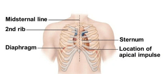
The heart is somewhat pyramid-shaped and lies within the pericardium in the middle mediastinum. The heart is situated at the center of the ‘chest’ and points ‘slightly towards left’. It is positioned posteriorly to the body of the sternum with one-third situated on the right and two-third on the left of the midline. The left ventricle involves the formation of the apex of the heart. It lies at the level of the fifth left intercostal space, 3.5 in. (9cm) from the midline.
Heart pumps
The heart pumps around 7,200 liters of blood in a day throughput body. On average, the heart beats about 100,000 times a day, i.e., around 3 billion eats in a lifetime. An adult’s heart beats about 60 to 80 times per minute, and a newborn baby’s heart beats faster than an adult which is about 70 to 190 beats per minute.
Weight of heart
The heart measures 12*8.5cm and weighs 310g (males) and 255g (females).
Connections
The broad connections of the heart include,
- Superficially: Bifurcation of the main pulmonary trunk
- Anteriorly: The body of the sternum and the adjoining coastal cartilages; left lung, and pleura (APEX)
- Posteriorly: Oesophagus, descending thoracic aorta, hemiazygos veins, azygos, and thoracic duct
- Inferiorly: Diaphragm
- Laterally: lungs and pleura
Structure of the heart walls
The heart wall composes of three layers enclosed in the pericardium;
- Epicardium is the outer layer of the heart wall and develops by the visceral layer of the serous pericardium.
- The myocardium is the muscular middle layer of the wall of the heart. It consists of excitable tissues and the conducting system.
- Endocardium-A middle concentric layer

The rest of the heart composes mainly of the subepicardial and subendocardial layers.
Structure and function of the Heart
The heart is subdivided into right and left halves by septa, and a constriction subdivides each half of the heart into the two major cavities, the upper cavity being called the atrium, the lower the ventricle.
Heart chambers
The heart, therefore, consists of four chambers;
- Two atria
- Left atrium
- Right atrium
- Two ventricles
- Left ventricle
- Right ventricle
Atria– small, thin-walled chambers. They are the receiving chambers for blood returning to the heart from the circulation process. Atria propels the blood into the adjacent ventricles and receives blood from the left atrium; pulmonary veins and right atrium; superior and inferior vena cava. The right atrium pushes blood via the tricuspid valve into the right ventricle. The left atrium pumps blood with the help of the bicuspid (mitral) valve into the left ventricle.
Ventricles– Discharging chambers of the heart. They pump blood to the pulmonary trunk (right ventricle) and aorta (left ventricle). The right ventricle propels blood via the pulmonary semilunar valve into the pulmonary trunk that is to be oxygenated in the lungs. Blood coming back from the lungs drains into the left atrium via the four pulmonary veins. The left ventricle pumps blood with the help of the aortic semilunar valve into the ascending aorta to supply blood to the body. The right ventricle receives blood from the right atrium through the tricuspid valve. The wall of the left ventricle is quite thicker than the right ventricle but the structure of both is almost similar. The thick wall is necessary to pump oxygenated blood at high pressure through the systemic circulation.
Heart valves
The heart has four valves. All four valves of the heart have one functional purpose of allowing the forward flow of blood but avoiding the backward flow. Heart valves ensure unidirectional blood flow through the heart composed of an endocardium with a connective tissue core. Heart valves guard the outflow of each chamber.
Atrioventricular valves between the atria and ventricles;
- The tricuspid valve (right side of the heart)- separates the right atrium from the right ventricle.
- Mitral valve/ bicuspid valve (left side of the heart)- separates the left atrium from the left ventricle.
- Both prevent backflow into the atrium.
Semilunar valves are present in the outflow tracts of the ventricles chambers. They include,
- The aortic valve (left side of the heart)- separates the left ventricle from the aorta.
- The pulmonary valve (right side of the heart)-separates the right ventricle from the pulmonary arteries.
- Both prevent backflow after ventricular contraction.
Also read: Cardiovascular Diseases: Leading Cause of Deaths Globally