Breast cancer develops when cells in your breast multiply and divide uncontrollably, resulting in a lump of tissue known as tumors. However, cancer symptoms include feeling a lump in your breast or observing abnormalities in the skin of your breasts. Moreover, experience a change in the size of your breast. Early detection can detect by mammograms.
Breast cancer is a type of cancer that grows in the breast cell. However, breast cancer is the second most common cancer diagnosed in women, after skin cancer. This cancer can affect both men and women, although it affects women significantly more frequently.
Significant funding for breast cancer research and awareness has aided in the development of cancer diagnosis and therapy. Early identification is a new personalized approach to therapy and a better knowledge of the disease has all contributed to an improvement in cancer survival rates. However, a steady decline in the number of fatalities linked with the disease.
What is Breast Cancer?
Breast cancer begins in the tissue of your breast. However, cancer develops when cells in the breast mutate (change) and expand out of control resulting in a mass of tissue (tumor). Like other cancers, breast cancer can enter and expand into the tissue that surrounds your breast. It can also spread to other parts of your body resulting in the formation of additional tumors. When this happens, it’s called metastasis.

What are the Types of Breast Cancer?
Breast cancer comes in a variety of types, including:
Infiltrated Ductal Carcinoma:
This cancer begins in your breast milk ducts, breaks through the duct wall, and spreads to surrounding breast tissue. However, cancer of this type is the most common.
Infiltrated Lobular Carcinoma:
This type of cancer starts in the lobules of your breast (where breast milk is produced) and has spread to the surrounding tissue. This cancer accounts for 10% to 15% of all cancers in women.
Ductal Carcinoma in situ:
Ductal carcinoma in situ is also known as Stage 0 cancer. It can consider precancerous by some because the cells haven’t spread beyond your milk ducts. This is a very treatable condition. However, quick treatment can require to prevent cancer from spreading to other tissues and becoming aggressive.
Inflammatory Cancer:
Inflammatory cancer is rare. It looks like an infection. Inflammatory breast cancers are caused by redness, swelling, pitting, and dimpling of the breast skin. Obstructive cancer cells in their skin’s lymph vessels cause it.
Causes:
According to doctors, cancer begins when some cells begin to grow abnormally. However, these cells proliferate and grow more rapidly than healthy cells, resulting in a lump or mass. However, cancerous cells can spread to lymph nodes and other parts of your body.
Breast cancer is commonly caused by cells in the milk-producing ducts (invasive ductal carcinoma). However, cancer can occur in lobules, which are glandular tissue. Alternatively, it can grow in different cells or tissues within the breast (invasive lobular carcinoma).
According to research, hormonal, lifestyle, and environmental factors have all been linked to an increased risk of cancer. However, no one understands why some people with no risk factors get cancer and others with risk factors never do. Moreover, cancer can produce by a complicated interaction between your genetic makeup and your environment.
What are the indications and symptoms of breast cancer in its early stages?

Breast cancer signs and symptoms are different in all cases. However, this cancer can show up in a variety of ways, including:
- A small lump or tumor that feels like a pea
- A change in the size, shape, or curve of your breast
- A lump or thickening in or near your breast or underarm that persists throughout your menstrual cycle
- A change in the look or feel of your breast or nipple skin (dimpled, puckered, scaly, or inflamed)
- Your breast or nipple skin is getting red
- A hardened marble-like area under your skin
- A clear or blood-stained fluid discharge from your nipple
Some people are unaware that they have cancer. That is why mammograms should be done regularly.
Risk Factors:
- After the age of 50: Cancer risk rises with age, with the cases detected beyond the age of 50.
- Mutations in the DNA: Certain genes, such as BRCA1 and BRCA2, have inherited changes (mutations). Breast and ovarian cancer are more common in women who have inherited these genetic changes.
- Reproductive History: Early menstrual periods before the age of 12 and Menopause after the age of 55 expose women to hormones for prolonged periods, increasing their risk of cancer.
- Breast or ovarian cancer in the family: If a woman has a mother, sister, or daughter (first-degree relative) or many family members on either her mother’s or father’s side of the family who have had breast or ovarian cancer, her chance of cancer can increase. A woman’s risk can further increase if she has a first-degree male relative who has cancer.
- Previous Radiation therapy: Women who received radiation therapy to the chest or breasts before the age of 30 have a greater risk of developing cancer later in life.
- Dense breast: As dense breasts have more connective tissue than fatty tissue, cancers can be difficult to spot on mammography. This cancer is more likely to develop in women who have big breasts.
- Non-cancerous breast disorders: Women who have had cancer before are more likely to have it again. Non-cancerous illnesses such as atypical hyperplasia and lobular carcinoma in situ have been associated with a higher risk of cancer.
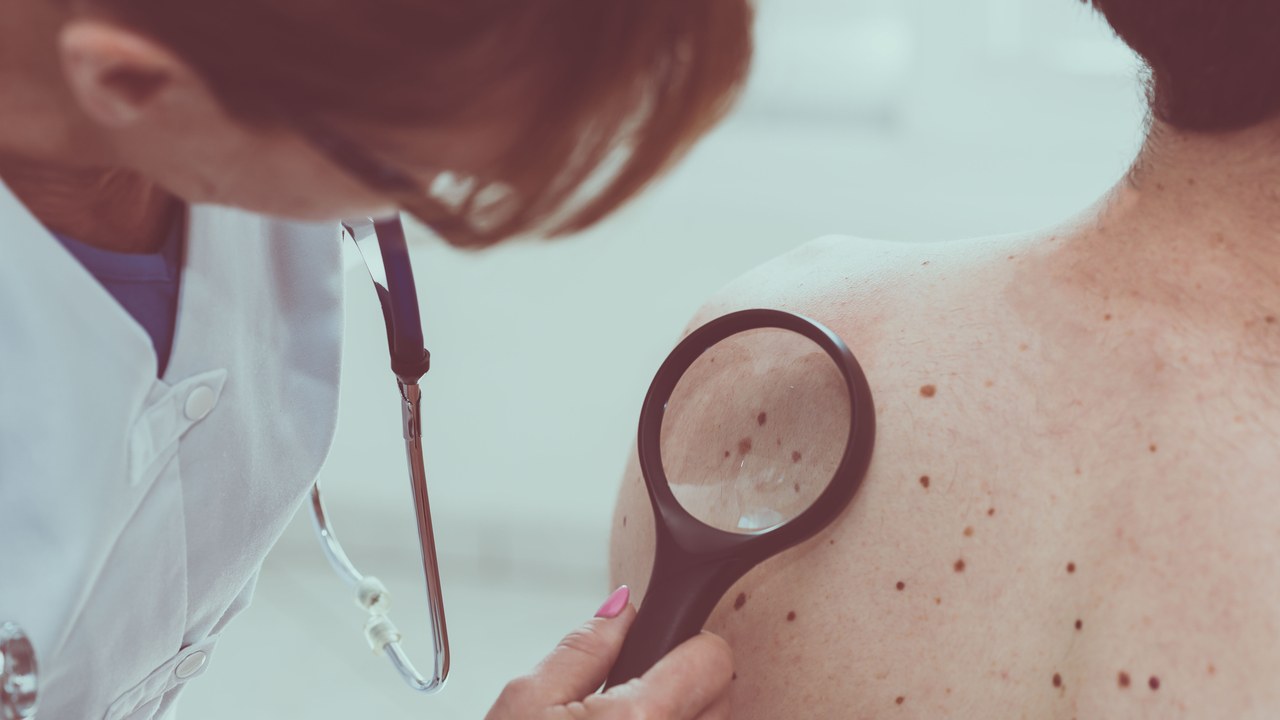
How is Breast Cancer Diagnosed?
A breast examination will perform by your healthcare provider. He will also inquire about your family history, medical history, and any symptoms. Tests to look for breast abnormalities will recommend by your health professional. These tests may involve the following:
- Mammogram:
Changes or abnormal growths in your breast may detect with these special X-ray scans. However, this cancer prevention usually includes the use of a mammogram.
- Ultrasonography:
Sound waves can use to take pictures of the tissues inside your breast during this test. It can use to detect lumps or abnormalities in the breast.
- PET scanning:
PET scans use unique dyes to highlight suspicious areas. Your health professional injects a special dye into your veins and uses a scanner to capture images.
- Magnetic resonance imaging:
MRI is a test that combines magnets and radio waves to create detailed images of the structures inside your breast.
Your healthcare provider may take a biopsy of your breast tissue if the imaging tests show anything abnormal. The sample will send to a pathology lab for analysis.